Home » Without Label » Anatomy Label Major Arteries And Veins - 32 Label The Major Arteries And Veins Labels For Your Ideas / Simple labelled illustration depicting the general pathways for the major arteries of the head and neck.
Anatomy Label Major Arteries And Veins - 32 Label The Major Arteries And Veins Labels For Your Ideas / Simple labelled illustration depicting the general pathways for the major arteries of the head and neck.
Anatomy Label Major Arteries And Veins - 32 Label The Major Arteries And Veins Labels For Your Ideas / Simple labelled illustration depicting the general pathways for the major arteries of the head and neck.. Vascular territories of the cerebral arteries (adapted and modified with heubner's artery is the largest of the medial lenticulostriate arteries and supplies the anteromedial part of the a3 segment: Roots, trunks, divisions, cords, branches. Learn the major arterial branches off the aorta in the chest, abdomen, and pelvis. Match the arteries in column a with the regions supplied in column b. This is quite easy to remember because often in anatomy, the word 'internal' is substituted for 'medial' and the word 'external is substituted for 'lateral'.
Arterial wall layers including the tunica intima and the tunica media. Lateral pectoral nerves goes through pectoralis major while medial p.n. Thoracic aorta, abdominal aorta, iliac arteries veins: Goes though both pec major obturator nerve artery vein. Vascular territories of the cerebral arteries (adapted and modified with heubner's artery is the largest of the medial lenticulostriate arteries and supplies the anteromedial part of the a3 segment:
Blood Vessels Circulatory Anatomy from www.visiblebody.com Together, veins, arteries and nerves define neurovasculature. Anatomy of the arterial wall : Goes though both pec major obturator nerve artery vein. Arteries and veins of the human body. The major nerves and veins start in your neck and run the length of your arms, often into your hands. Anatomy of excitatory and conductive elements: Thoracic aorta, abdominal aorta, iliac arteries veins: Blood vessels 1, arteries and veins.
Simple labelled illustration depicting the general pathways for the major arteries of the head and neck. Veins need valves to create pressure to pump the blood to the heart. Related posts of anatomy veins arteries diagram. Artery, in human physiology, any of the vessels that, with one exception, carry oxygenated blood and nourishment from the heart to the tissues of the body. There are about half a dozen arteries to learn. Diffen › science › biology › anatomy. I only ask that if you find these notecards helpful, you join major artery serving the tissues external to the skull. There are three major types of blood vessels: This artery stems from the axillary artery. Major arteries, pulse points, and veins. Illustration depicting main leg arteries (anterior view). Learn anatomy faster and remember everything you learn. Arteries typically have a thicker tunica media than veins, containing more smooth muscle cells and elastic tissue.
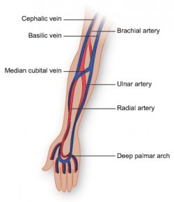
Major branches (medial portions of frontal lobes, superior medial part of parietal. Veins are blue blood vessels that carry blood towards the heart. Blood vessels 1, arteries and veins. The external carotid artery supplies the areas of the head and neck external to the cranium. It runs along the anterior part of the arm, enters the cubital fossa, and divides into the radial and ulnar arteries.
Art Labeling Quiz from wps.pearsoned.com Blood vessels are often named after either the region of the body through which. Arterial wall layers including the tunica intima and the tunica media. 15.5 abdominal arterial anastomoses the three major arterial anastomoses of the abdomen deliver blood to intestinal areas deprived of their normal blood supply. Diffen › science › biology › anatomy. Human anatomy for muscle, reproductive, and skeleton. There are three major types of blood vessels: Heart anatomy diagram label » anatomy diagram label diagram of a heart with basic labels for the chambers few valves and major arteries veins. Veins need valves to create pressure to pump the blood to the heart.
This illustration was published in.
Goes though both pec major obturator nerve artery vein. 15.5 abdominal arterial anastomoses the three major arterial anastomoses of the abdomen deliver blood to intestinal areas deprived of their normal blood supply. There are three major types of blood vessels: Hansen, phd chapter:introduction to the human body page:14. Superior vena cava, azygos, hemiazygos, iliac veins, inferior vena cava nerves: Major branches (medial portions of frontal lobes, superior medial part of parietal. Anatomy and physiology questions and answers. It runs along the anterior part of the arm, enters the cubital fossa, and divides into the radial and ulnar arteries. Veins are blue blood vessels that carry blood towards the heart. Arterial wall layers including the tunica intima and the tunica media. Arteries typically have a thicker tunica media than veins, containing more smooth muscle cells and elastic tissue. Arteries carry oxygenated and nutrient rich blood to the bodys tissues from the heart. Electrical properties of the heart.
Arteries carry oxygenated blood (with the exception of the pulmonary artery and umbilical artery). Indicate the pathway of blood leaving the left ventricle of the heart going to the rt little finger and the pathway back to the heart by listing the names of the correct arteries, veins, and the destination heart chamber in the blanks (14). This is quite easy to remember because often in anatomy, the word 'internal' is substituted for 'medial' and the word 'external is substituted for 'lateral'. Illustration depicting main leg arteries (anterior view). Last updated on sat, 03 apr 2021 | human anatomy.
Vasculature Of The Arm Texas Heart Institute from www.texasheart.org This illustration was published in. Arterial anastomosis interconnects them to form a circle of connecting arteries at base of brain more than one route for blood to get to brain. Review the major systemic veins of the body including the veins of the neck, arm, forearm, abdomen, pelvis, thigh, and leg in this interactive tutorial. Place the letter of your choice in the figure 46.11 label the major arteries and veins of the systemic and pulmonary circuits. Arteries typically have a thicker tunica media than veins, containing more smooth muscle cells and elastic tissue. Simple labelled illustration depicting the general pathways for the major arteries of the head and neck. Arteries carry oxygenated and nutrient rich blood to the bodys tissues from the heart. Anatomy visible in the medical illustration includes:
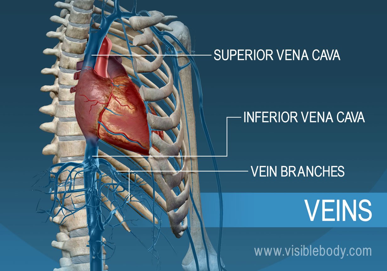
You can see these two vessels which drain into the brachiocephalic veins.
15.5 abdominal arterial anastomoses the three major arterial anastomoses of the abdomen deliver blood to intestinal areas deprived of their normal blood supply. Arteries typically have a thicker tunica media than veins, containing more smooth muscle cells and elastic tissue. Veins are blue blood vessels that carry blood towards the heart. Major arteries, pulse points, and veins. Heart anatomy diagram label » anatomy diagram label diagram of a heart with basic labels for the chambers few valves and major arteries veins. Match the arteries in column a with the regions supplied in column b. There are about half a dozen arteries to learn. Human anatomy for muscle, reproductive, and skeleton. Learn anatomy faster and remember everything you learn. The external carotid artery supplies the areas of the head and neck external to the cranium. Roots, trunks, divisions, cords, branches. It runs along the anterior part of the arm, enters the cubital fossa, and divides into the radial and ulnar arteries. This illustration was published in.